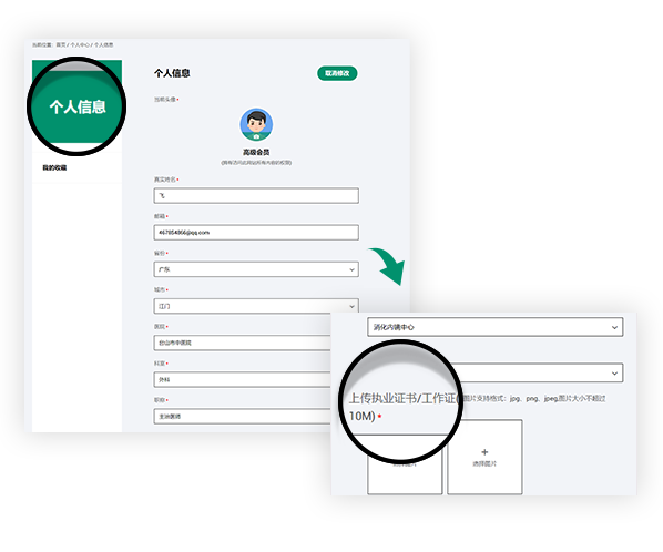
Join us you will be able to get the following rights
Get fresh academic and clinical information
Sign up for exclusive endoscopy contests and training courses
Use online training software
Watch the LIVE of academic conferences and surgery
Linked color imaging can enhance recognition of early gastric cancer by high color contrast to surrounding gastric intestinal metaplasia
Linked color imaging can enhance recognition of early gastric cancer by high color contrast to surrounding gastric intestinal metaplasia
H Fukuda, Y Miura, H Osawa, T Takezawa… - Journal of Gastroenterology, 2019
Background: Linked color imaging (LCI) increases the visibility of early gastric cancers, which may be associated with characteristic findings including background purple mucosae. These lesions are found in areas of chronic gastritis and surrounding mucosa. The aim of this study is to objectively characterize these lesions by color differences and color component values using LCI.
Methods: Fifty-two patients with early gastric cancer were enrolled. Color differences were calculated prospectively in malignant lesions and adjacent mucosa and compared with histological findings in resected specimens. Color component values of L*, a*, and b* were compared between purple and non-purple mucosae in areas of chronic gastritis. Based on histological findings, the accuracy of identifying gastric intestinal metaplasia was calculated.
Results: Cancers and surrounding mucosa in 74% of lesions had similar colors using white light imaging (WLI), whereas purple mucosa surrounded part or all of cancers appearing orange–red, orange or orange–white using LCI. Greater color differences were seen using LCI compared to WLI, including flat-type cancers, leading to higher contrast. The surrounding purple mucosa corresponded histologically to intestinal metaplasia, facilitating the identification of malignant lesions. Forty lesions (83%) with purple mucosa and eight lesions (17%) with non-purple mucosa in areas of chronic gastritis were diagnosed as intestinal metaplasia by biopsy (83% accuracy). Color component values of purple mucosa differ significantly from those of non-purple mucosae.
Conclusions: LCI images have higher color contrast between early gastric cancers and surrounding mucosa compared to WLI. A characteristic purple color around gastric cancers using LCI represents intestinal metaplasia.
声明
富士胶片内镜世界(LIFE World)所登载的内容及其版权和使用权归作者本人与富士胶片所有。如发现会员擅自复制、更改、公开发表或其他以盈利为目的的使用,富士胶片将追究其法律责任。网站信息中涉及的治疗手技皆为术者个人针对该名患者特定体质及健康状况所采取的手法;术者对器械和药品种类的选择,也受到手术发生时间、地点等诸多因素的影响。因而相关内容及信息仅供会员参考。如盲目使用网站信息中涉及的治疗手技而发生意外,恕富士胶片及本网站对此不承担任何责任。


