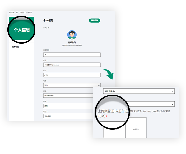
Join us you will be able to get the following rights
Get fresh academic and clinical information
Sign up for exclusive endoscopy contests and training courses
Use online training software
Watch the LIVE of academic conferences and surgery
Clinical significance and influencing factors of linked color imaging technique in real-time diagnosis of active Helicobacter pylori infection
Clinical significance and influencing factors of linked color imaging technique in real-time diagnosis of active Helicobacter pylori infection
L Wang, XC Lin, HL Li, XS Yang, L Zhang… - Chinese Medical Journal, 2019
Background: Determining the Helicobacter pylori (H. pylori) infection state during the gastroscopic process is important but still challenging. The linked color imaging (LCI) technique might emphasize the mucosal color change after H. pylori infection, which might help the diagnosis. In the present study, we aimed to compare the LCI technique with traditional white light imaging (WLI) endoscopy for diagnosing active H. pylori infection.
Methods: We collected and analyzed gastroscopic images from 103 patients in our hospital from November 2017 to March 2018, including both LCI and WLI modes. All images were randomly disordered and independently evaluated by four endoscopists who were blinded to the H. pylori status of patients. In addition, the H. pylori state was determined by both rapid urease test and pathology staining. The sensitivity, specificity, positive prediction value (PPV), and negative prediction value (NPV) were calculated for the detection of H. pylori infection. Moreover, the kappa value and interclass correlation coefficient (ICC) were used to evaluate the inter-observer variety by SPSS 24.0 software.
Results: Of the 103 enrolled patients, 27 of them were positive for H. pylori infection, while the 76 patients were negative. In total, 388 endoscopic images were selected, including 197 WLI and 191 LCI. The accuracy rate for H. pylori evaluation in the corpus LCI group was significantly higher than other groups (81.2% vs. 64.3%–76.5%, χ2 = 34.852, P < 0.001). Moreover, the corpus LCI group had the optimal diagnostic power with the sensitivity of 85.41% (95% confidence interval [CI]: 76.40%–91.51%), the specificity of 79.71% (95% CI: 74.38%–84.19%), the PPV of 59.42% (95% CI: 50.72%–67.59%), and the NPV of 94.02% (95% CI: 89.95%–96.56%), respectively. The kappa values between different endoscopists were higher with LCI than with WLI (0.433–0.554 vs. 0.331–0.554). Consistently, the ICC value was also higher with LCI than with WLI (0.501 [95% CI: 0.429–0.574] vs. 0.397 [95% CI: 0.323–0.474]). We further analyzed the factors that might lead to misjudgment, revealing that active inflammation might disturb WLI judgment (accuracy rate: 58.70% vs. 76.16%, χ2 = 21.373, P < 0.001). Atrophy and intestinal metaplasia might affect the accuracy of the LCI results (accuracy rate: 66.96% vs. 73.47%, χ2 = 2.027; 68.42% vs. 73.53%, χ2 = 1.594, respectively); however, without statistical significance (P = 0.154 and 0.207, respectively).
Conclusions: The application of LCI at the corpus to identify H. pylori infection is reliable and superior to WLI. The inter-observer variability is lower with LCI than with WLI.
声明
富士胶片内镜世界(LIFE World)所登载的内容及其版权和使用权归作者本人与富士胶片所有。如发现会员擅自复制、更改、公开发表或其他以盈利为目的的使用,富士胶片将追究其法律责任。网站信息中涉及的治疗手技皆为术者个人针对该名患者特定体质及健康状况所采取的手法;术者对器械和药品种类的选择,也受到手术发生时间、地点等诸多因素的影响。因而相关内容及信息仅供会员参考。如盲目使用网站信息中涉及的治疗手技而发生意外,恕富士胶片及本网站对此不承担任何责任。


