Member login
会员登录返回登录
微信扫一扫登录
扫描成功请在微信中点击确认即可登录
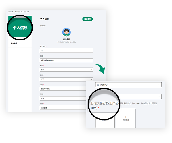
会员绑定
Register member
Join us you will be able to get the following rights
Get fresh academic and clinical information
Sign up for exclusive endoscopy contests and training courses
Use online training software
Watch the LIVE of academic conferences and surgery


登录

Linked color imaging enhances endoscopic detection of sessile serrated adenoma/polyps
收藏已收藏
0
774
LCI文献
Linked color imaging enhances endoscopic detection of sessile serrated adenoma/polyps
2021/09/06LCI文献
收藏已收藏
0
774
查看全文
D Fujimoto, N Muguruma, K Okamoto… - Endoscopy International Open, 2018
Background and study aims: Although new image-enhanced endoscopy (IEE) technologies such as blue laser imaging (BLI), BLI-bright, and linked color imaging (LCI) have been developed, their utility for the detection of sessile serrated adenoma/polyps (SSA/Ps) is still unclear. This study aimed to evaluate the utility of BLI, BLI-bright, and LCI for SSA/P detection in still image examinations and in a prospective randomized controlled trial (RCT).
Patients and methods: A group of 6 expert and non-expert endoscopists read 200 endoscopic still images containing SSA/P lesions using white light image (WLI), BLI, BLI-bright, and LCI. Color differences were calculated using the color space method. A prospective RCT of tandem colonoscopy with WLI and LCI was performed. Patients with SSA/P and those with a history of SSA/P that had been endoscopically removed were enrolled and randomly allocated to WLI-LCI or LCI-WLI groups. Additional endoscopic detection rates for SSA/P were compared between the 2 groups.
Results: LCI showed the highest SSA/P detection rate among the 4 modes for both expert and non-expert endoscopists. The detection rate with LCI for the 6 expert endoscopists (mean 98.3 ± standard deviation 2.0 %) was significantly higher than that with WLI (86.7 ± 6.0 %, P < 0.01). Likewise, the detection rate with LCI for the 6 non-expert endoscopists (92.3 ± 2.9 %) was significantly higher than that with WLI (72.7 ± 11.5 %, P < 0.01). The color difference of SSA/P with LCI was the highest among the 4 modes, and was significantly higher than with WLI (median 15.9, (interquartile range 13.7 – 20.6) vs. 10.2, (7.6 – 14.2); P < 0.0001). In the RCT, a total of 44 patients (WLI-LCI 22 vs. LCI-WLI 22) underwent colonoscopy. The additional detection rate for SSA/P in the second inspection in the WLI-LCI group (21.6 %, 8/37) was significantly higher than in the LCI-WLI group (3.2 %, 1/31; P = 0.02). The small, flat, non-mucus and isochromatic SSA/Ps in the transverse colon were detected more frequently in the second inspection with LCI.
Conclusions: LCI was the most sensitive mode for SSA/P detection among WLI, BLI, BLI-bright, and LCI in the still image examinations. Our RCT strongly suggests that LCI is superior to conventional WLI for SSA/P detection during colonoscopy.
声明
富士胶片内镜世界(LIFE World)所登载的内容及其版权和使用权归作者本人与富士胶片所有。如发现会员擅自复制、更改、公开发表或其他以盈利为目的的使用,富士胶片将追究其法律责任。网站信息中涉及的治疗手技皆为术者个人针对该名患者特定体质及健康状况所采取的手法;术者对器械和药品种类的选择,也受到手术发生时间、地点等诸多因素的影响。因而相关内容及信息仅供会员参考。如盲目使用网站信息中涉及的治疗手技而发生意外,恕富士胶片及本网站对此不承担任何责任。

最新留言