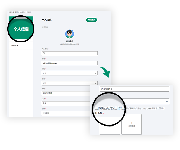
Join us you will be able to get the following rights
Get fresh academic and clinical information
Sign up for exclusive endoscopy contests and training courses
Use online training software
Watch the LIVE of academic conferences and surgery
联动成像技术提高了内镜下结肠非颗粒状扁平病变的可见性
联动成像技术提高了内镜下结肠非颗粒状扁平病变的可见性
联动成像技术提高了内镜下结肠非颗粒状扁平病变的可见性
Linked-color imaging improves endoscopic visibility of colorectal nongranular flat lesions
T Suzuki, T Hara, Y Kitagawa, H Takashiro… - Gastrointestinal Endoscopy, 2017
本研究结果提示LCI提高了结肠扁平病变的可见性,有助于提高这些病变的检出率。
Conclusions: The present findings suggest that LCI increases the visibility of colorectal flat lesions and contributes to improvement of the detection rate for these lesions.
01
背景与目的
作为一种新的图像增强内镜(IEE)技术,联动成像技术(Link Color Imaging, LCI)提供增强色彩的明亮图像。为了提高难以检测的结直肠扁平病变的检出率,我们从可见性的角度探讨了LCI的有效性。
Background and study aims: As a newly developed image-enhanced endoscopy (IEE) technique, linked-color imaging (LCI) provides very bright images with enhanced color tones. With the objective of improving the detection rate of colorectal flat tumor lesions, which are difficult to detect, we examined the usefulness of LCI from the viewpoint of visibility.
02
方法
本研究采用53例连续的非颗粒扁平肿瘤。内镜下图像采用白光成像(WLI)、蓝光成像(BLI)-bright和LCI模式。对于每一个病灶,我们选择了通过WLI、 BLI-bright和LCI模式获得的各1张图像。 六名内镜医生解读这些图像。 通过使用以前报告的可见性等级,我们在1到4的等级上对可见性打分。
Methods: Fifty-three consecutive nongranular flat tumors were used in this study. Endoscopic images were acquired by white-light imaging (WLI), blue-laser imaging (BLI)-bright, and LCI modes. For each lesion, we selected 1 image each acquired by WLI, BLI-bright, and LCI modes. Six endoscopists interpreted the images. By using a previously reported visibility scale, we scored the visibility level on a scale of 1 to 4.
03
结果
可见性评分均值(±标准差):WLI为2.74±1.08,BLI-bright为2.94±0.97,LCI为3.36±0.72。BLI-bright的得分显著高于白光组(P < .001),而LCI的得分又高于BLI-bright (P< .001)。 当我们将专家与实习医生进行比较时,专家的相应得分分别为2.83±1.06、3.17±0.88、3.40±0.74,与所有内镜医师的得分趋势相似。对于实习医生而言, WLI(2.65±1.10)与BLI-bright(2.71±1.00)无显著性差异,但LCI(3.31±0.69)显著高于WLI和BLI-bright (P < 0.001)。 当只分析无蒂锯齿状腺瘤/息肉病变时,LCI显著高于其他2组。
Results: The mean (± standard deviation) visibility scores were 2.74 ± 1.08 for WLI, 2.94 ± 0.97 for BLI-bright, and 3.36 ± 0.72 for LCI. The score was significantly higher for BLI-bright compared with WLI (P < .001) and again higher for LCI compared with BLI-bright (P < .001). When we compared between experts and trainees, the corresponding scores of experts were 2.83 ± 1.06, 3.17 ± 0.88, and 3.40 ± 0.74, with a tendency similar to the scores of all endoscopists. For the trainees, there was no difference between the scores for WLI (2.65 ± 1.10) and BLI-bright (2.71 ± 1.00), but the score for LCI (3.31 ± 0.69) was significantly higher than that for WLI or BLI-bright (P < .001). When only sessile serrated adenoma/polyp lesions were analyzed, LCI remained significantly higher than the other 2.
声明
富士胶片内镜世界(LIFE World)所登载的内容及其版权和使用权归作者本人与富士胶片所有。如发现会员擅自复制、更改、公开发表或其他以盈利为目的的使用,富士胶片将追究其法律责任。网站信息中涉及的治疗手技皆为术者个人针对该名患者特定体质及健康状况所采取的手法;术者对器械和药品种类的选择,也受到手术发生时间、地点等诸多因素的影响。因而相关内容及信息仅供会员参考。如盲目使用网站信息中涉及的治疗手技而发生意外,恕富士胶片及本网站对此不承担任何责任。


