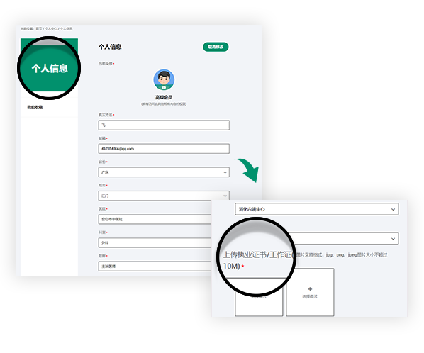
Join us you will be able to get the following rights
Get fresh academic and clinical information
Sign up for exclusive endoscopy contests and training courses
Use online training software
Watch the LIVE of academic conferences and surgery
基于内镜的联动成像技术和病理学评估胃肠上皮化生
基于内镜的联动成像技术和病理学评估胃肠上皮化生
基于内镜的联动成像技术和病理学评估胃肠上皮化生
Gastric intestinal metaplasia assessment between linked color imaging based on endoscopy and pathology
G Zhang, J Zheng, L Zheng, S Yu, C Jiang… - Scandinavian Journal of Gastroenterology, 2021
我们的研究证明了EGGIM诊断肠化生程度的能力,并表明EGGIM与OLGIM分期相关。EGGIM 4期是识别OLGIM III/IV患者的最佳分界值。
Conclusions: Our findings demonstrated the ability of EGGIM for diagnosing the extent of intestinal metaplasia and showed that EGGIM is related to OLGIM staging. EGGIM of 4 was the best cut-off value for identifying OLGIM III/IV patients.
01
目的
累计证据表明,联动成像技术(LCI)可用于识别胃肠上皮化生(GIM)。我们的目标是使用LCI开发GIM的内镜分级(EGGIM)
Objective: Cumulative evidence suggests that linked color imaging (LCI) can be used to identify gastric intestinal metaplasia (GIM). We aimed to develop endoscopic grading for GIM (EGGIM) with LCI.
02
方法
招募277名接受高分辨率白光胃镜检查的患者,再用LCI模式检查以进行EGGIM评估。用LCI模式进行整个胃黏膜检查。记录胃窦、胃体的小弯和大弯以及切迹五个区域的图像。对于每个区域,0分(无GIM)、1分(局部GIM,≤30%的区域)和2分(广泛GIM,>30%的区域)归纳为10期。如果根据内镜检查结果怀疑GIM,则进行有针对性的活检;如果GIM不明显,则根据悉尼系统进行随机活检以评估基于肠化的胃炎评价(OLGIM)。
Methods: Two hundred and seventy-seven patients who underwent high-resolution white-light gastroscopy followed by LCI for EGGIM estimation were included. LCI was performed for the entire mucosa, and images of five areas each were recorded from the lesser and greater curvatures of the antrum and corpus, and for the incisura. For each area, scores of 0 (no GIM), 1 (focal GIM, ≤30% of the area), and 2 (extensive GIM, >30% of the area) were attributed for 10 points. If GIM was suspected based on endoscopy findings, targeted biopsies were performed; if GIM was not evident, random biopsies were performed according to the Sydney system to estimate the operative link on GIM (OLGIM).
03
结果
分别有136、70、37、28和 6名患者的GIM评估为OLGIM 0、I、 II、 III和IV。OLGIM III/IV诊断,受试者工作特性曲线下面积为0.949(95% CI 80.3%–99.3%)。EGGIM 4期的敏感性和特异性分别为94.12% (95% CI 80.3%–99.3%)和 86.42% (95% CI 81.5%–90.5%),被 确认为识别OLGIM III/IV患者的最佳分界值。
Results: GIM was staged as OLGIM 0, I, II, III, and IV in 136, 70, 37, 28, and 6 patients, respectively. For OLGIM III/IV diagnosis, the area under the receiver operating curve was 0.949 (95% CI 0.916–0.972). EGGIM of 4, with sensitivity and specificity of 94.12% (95% CI 80.3%–99.3%) and 86.42% (95% CI 81.5%–90.5%), respectively, was determined the best cut-off value for identifying OLGIM III/IV patients.
声明
富士胶片内镜世界(LIFE World)所登载的内容及其版权和使用权归作者本人与富士胶片所有。如发现会员擅自复制、更改、公开发表或其他以盈利为目的的使用,富士胶片将追究其法律责任。网站信息中涉及的治疗手技皆为术者个人针对该名患者特定体质及健康状况所采取的手法;术者对器械和药品种类的选择,也受到手术发生时间、地点等诸多因素的影响。因而相关内容及信息仅供会员参考。如盲目使用网站信息中涉及的治疗手技而发生意外,恕富士胶片及本网站对此不承担任何责任。


